
Automated Model-Based Quantitative Analysis of FDG PET Phantoms
Quality control plays an increasingly important role in quantitative PET imaging and is typically performed using phantoms. To simplify the quantitative analysis process, we have developed a fully automated algorithm (Fig. 1) for two common PET/CT quality assurance phantoms: the NEMA NU-2 IQ and SNMMI/CTN oncology phantom. Moreover, our approach can easily be adapted to other phantoms that utilize spherical inserts by utilizing simple phantom model descriptions. We believe that the developed algorithm/software provides value to the PET imaging community. Therefore, we are actively working on developing a dissemination strategy. Specifically, we are considering: a) dissemination through a society and b) making the source code available amongst other avenues, including the publication of the algorithm in form of a paper. Updates regarding the availability of our approach will be posted here in the near future.
Since October of 2019, an expanded version of our software was made available via the Society of Nuclear Medicine & Molecular Imaging.
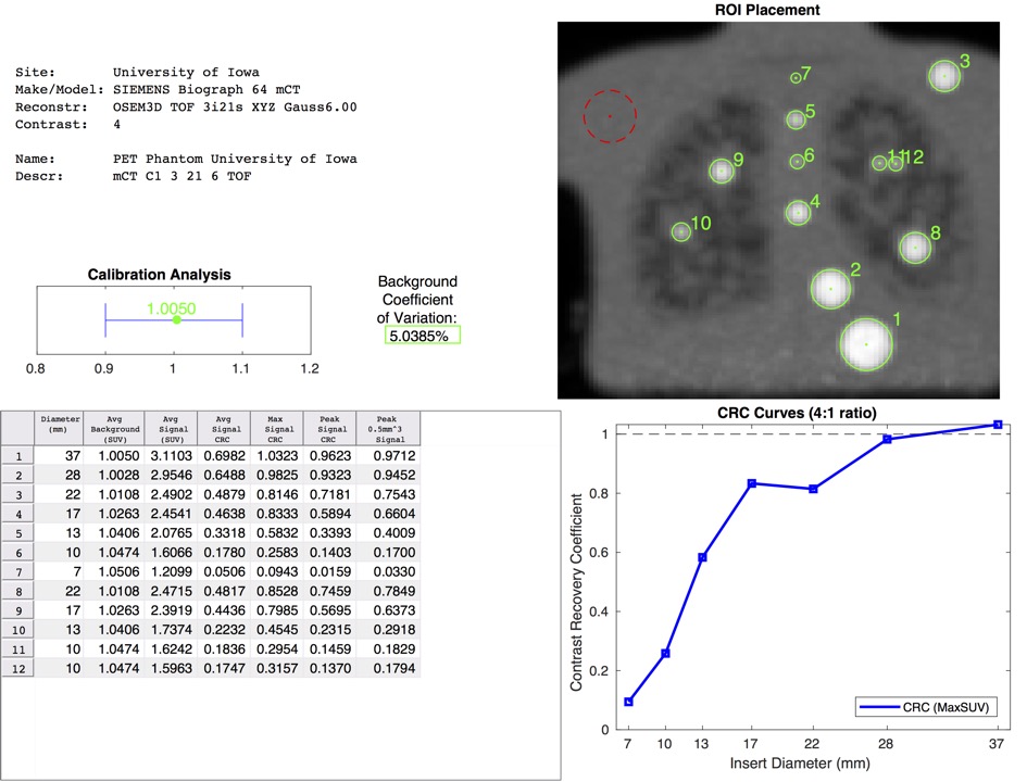
Fig. 1. Example of an automatically generated PET phantom analysis report.
Related Work
Note that for the analysis of PET cylinder phantoms, we have developed the following extension for 3D Slicer. PET Cylinder Phantom Analysis
The PET Phantom Analysis Extension enables automated analysis of Uniform Cylinder Phantoms in PET scans utilized for quality control purposes.
Source code: https://github.com/QIICR/SlicerPETPhantomAnalysis
Documentation: https://www.slicer.org/wiki/Documentation/Nightly/Extensions/PETPhantomAnalysis
Version history: Available since Slicer 4.10.0.
Functionality verified up to Slicer 5.0.2.
Acknowledgements
This work was supported in part by NIH NCI QIN program grant U01 CA140206.
Contact
The contents of this website does not necessarily represent the official views of the NIH/NCI.